Aristotle said it first, the whole is greater than the sum of the parts. Christian Smutz brought the concept of holism to the modern era with his book Holism and Revolution. This background was highlighted in greater detail in my previous post on Wholistic vs Holistic Cancer Treatment.
The general concept is that the whole transcends the individual parts. Holistic medicine is a non-compartmentalized approach to healthcare. With less information, the ancients were more holistic in their approach than the experts of today, who have significantly more information.
A holistic approach to cancer is no different. A holistic approach to cancer is found in causation, testing, treatment, and in maintenance therapy. It is holistic in the whole sense of the word. This post will look at a holistic approach to some of the causes of cancer.
A holistic approach to the causes of cancer includes:
- Lifestyle
- Epigenetics
- Hypoxia
- Inflammation
- Mitochondria
- Metabolic dysfunction
- Cell and mitochondrial Voltage
- Acid/Base balance
- Reduction/oxidation balance (Redox)
- Tumor Microenvironment (TME)
- Immune dysfunction
- Hormones
- Toxicants and detoxification dysfunction
- Infection
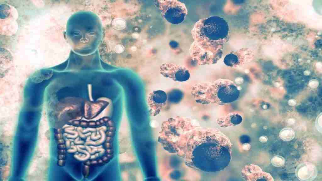
- The Gut
- And other associated deficiencies
There are and can be many more contributors, but this post is going to be long enough. To condense the quantity of information in this post, I will break it up into a series of posts. I want to highlight each point in detail individually.
Lifestyle
Conventional medicine continues to push denial of the connection between lifestyle, particularly diet, and cancer. The science is precise on the matter. In contrast to the opinion often espoused by physicians, including conventional Oncologists, we are products of our collective environment, and our lifestyle choices dictate much of our cellular environment. No matter the denial, the evidence points to the contrary. What we put in our mouths affects cancer risk and cancer healing.
Our nutrition choices, or lack thereof, can benefit the whole of the body or damage the whole of the body. Lifestyle can include diet, stress, sleep/wake cycles, relationships, exercise/activity level, weight, home, and many others. A recently published article highlighted the connection between diet and cancer [i]. In this study model, dietary choices increase the toxicity of chemotherapy up to 100-fold. The mechanism, by researchers at the University of Virginia, was found to be through the dietary alterations in the gut microbiome. I have often said that diet is love language, or not, with our DNA. This study shows that diet is the means to alter the gut microbiome to increase or possibly decrease chemotherapy toxicity. In essence, the lifestyle choice of what we put in our mouth lays the groundwork for toxicity from chemotherapy. So much for the idea that nutrition plays no role in cancer treatment.
In fact, If doctors are telling patients with cancer to eat whatever they want, i.e., hamburgers, steak, and sugar... they are in effect, increasing the toxicity and morbidity of treatment in patients. There is an important phrase that should be on the tongue of every physician—“first do no harm”. This study just focused on diet. This point doesn’t take into account all the other individual lifestyle choices. Lifestyle choices do not exist in a bubble but instead exist as a collective combination to impact our body for health or for dis-ease. They are our choices.
Epigenetics
Epigenetics is an exciting new science that is making tremendous progress and promise in the treatment of dis-eases like cancer.
Epigenetics means "above genetics". The epigenome is the total number of modifications of the DNA (genome) in response to the environment (i.e., diet, stress...). This is the body's attempt to regulate the activity and expression of all the genes (epigenetics) within the genome in response to its environment. While DNA is the different pieces of the puzzle that is the body, epigenetics is the directions for how those genes, the "pieces," are used in response to the environment. This process of adaptation occurs throughout life. These adaptations are inheritable. These adaptations can be passed from generation to generation for generations. This inheritable epigenetic modifications is called Transgenerational Inheritance [ii]. Epigenetic alterations can and will occur at any time, including conception, pregnancy, puberty, stress, and beyond. These are normal processes that take place throughout this existence we call life and control how a body changes and adapts to its environment. In addition to these normal processes, it is apparent that environmental factors and life events can affect each individual's epigenome as well, which means you have a significant amount of control over your health now and your health potential in the future. Epigenetics brings each individual’s healing potential within their grasp.
Scientists are evaluating and testing new therapies that will use the epigenome to fight cancer. Just look at chemotherapy alone. Maximum tolerated chemotherapy is associated with significant, serious side effects. Instead of using high doses of toxic chemotherapy to attack the cancer cells, patients in trials are receiving lower doses of medication as a result of epigenomic targeting, explicitly directed towards the cancer cells while bypassing healthy cells [iii]. These patients are showing positive results and are suffering from fewer side effects from the treatments. Now there is a novel concept, target the cancer and not the patient.
Hypoxia
Oxygen is essential to life on earth. Hypoxia is the lack of oxygen. Without oxygen, life in its current form would not exist. Hypoxia is present at the genesis stage of cancer—called carcinogenesis [iv] [v]. As critical as oxygen is to life, the absence of oxygen is equally essential to the development of cancer. The whole body cannot be hypoxic. This would equal no life.
Hypoxia is critical to the Tumor Microenvironment (TME). The TME specifically will be discussed later in this series. Hypoxia induces signaling, such as Hypoxia-Inducible Factor-1alpha, to promote and propagate cancer [vi]. Hypoxia in the TME alters energy production via the Warburg effect [vii]. The hypoxic effect increases lactate production to increase the acidic environment that is so characteristic of the cancer TME [viii] [ix] [x]. In addition, hypoxia alters the immune system in the TME to protect and preserve the growing tumor from the immune system [xi] [xii], all at the expense of the body. Hypoxia even promotes cancer treatment resistance to conventional radiation [xiii] and chemotherapy [xiv]. Beyond the driving force that is hypoxia in the genesis of cancer, hypoxia is the major push behind the spread of cancer via metastasis [xv] [xvi] [xvii]. Hypoxia drives the production of blood vessel growth (angiogenesis) [xviii].[xix] that is so critical to the physical cell escape in metastasis. Hypoxia is also essential in how cancer can escape the immune system, both locally and systemically, in the process of metastasis.
Inflammation
Inflammation is the bed that cancer lies in. Inflammation is not de-facto the enemy of the body and is an over-simplified view of inflammation. Inflammation is, in fact, a critical, necessary component of the healing process. Just look at a paper cut. Immediately, the site is painful, red, swollen, and hot. These are the cardinal signs and symptoms of inflammation that prevent secondary infection and initiate the healing process. However, in the described setting of the paper cut, the inflammation subsides once the threat of secondary infection is gone, and the healing process is in full motion. In cancer, inflammation does not turn off, but in fact, turns on the body. In many ways, cancer uses the immune system and inflammation signaling to co-opt the immune system against the body.
Cancer requires chronic inflammation, yet cancer stimulates the production of inflammation. Inflammation is a by-product of immune system signaling. Immune system signaling and inflammation can inhibit cancer initiation, growth, and spread. Likewise, Immune signaling and inflammation can promote cancer initiation, growth, and spread. In many ways, NF-kappaB sits at this crossroad of inflammation and cancer [xx]. NF-kappaB is a critical genetic transcription factor that stimulates inflammation in cancer to promote tumorigenesis [xxi] [xxii]. Disordered immune function and Inflammation are present in the Tumor Microenviroment (TME) [xxiii]. An Inflammatory TME promotes NF-kappaB activation. It is the activation of NF-kappaB that further stimulates the production of inflammatory signals, called cytokines, that also increase pro-carcinogenic inflammation and even immune suppression [xxiv] [xxv]. The result is dysfunction of the immune system, evasion of the immune system, and even suppression of the immune system, which leads to growth [xxvi], survival [xxvii], invasion and metastasis of cancer [xxviii].
Mitochondria
What is the meaning of life? Not from a spiritual perspective. Not from a psychological perspective, but rather, from a biochemistry perspective? Life is about energy—the energy to heal, energy to repair, energy to fight infection, and the energy to grow. The capacity to produce energy is at the core of whether life exists or not. A body that can make energy efficiently can do all the things that are required to survive and thrive. A body that can not produce energy does not thrive and does not survive. That same applies to the cell. After all, the body is estimated to contain 3.72 × 10(13) number of cells [xxix]. That is 3,720,000,000,000,000 or 3.72 quadrillion. But who is counting? More, a cell that cannot make energy, cannot survive, cannot thrive and is targeted for destruction and recycling through the normal biochemical processes of apoptosis and autophagy.
Mitochondria are at the core of this biochemical life perspective. Mitochondria are the energy powerhouses of the cell. Mitochondria exist within every cell because every cell requires energy production to perform the day to day tasks necessary to survive. They must co-exist. Without mitochondria, there is no sustainable capacity for a cell to make energy. The energy production pathways of glycolysis, the Krebs cycle, and the electron transport chain are the energy production pathway forward within every mitochondrion for the benefit for cell survival.
The question is, which came first, the chicken or the egg? Does mitochondrial dysfunction initiate cancer, or does the process of cancer initiate mitochondrial dysfunction? As in most cases, the answer is yes. Yes, mitochondrial dysfunction is involved in the genesis of cancer, and yes, cancer is involved in the genesis of mitochondrial dysfunction. Both are proven true [xxx]. They are not independently true, but they are simultaneously true. The process of the biochemistry of cancer can no longer be viewed through a linear, one-dimensional sequence of events. That thinking is compartmentalization. That thinking is the modus operandi of conventional medicine. That thinking is not holistic.
The scientific literature leaves little evidence for any other conclusion, but that cancer is the result of poor adaptation to metabolic stress in an attempt to survive. I want you to look at cancer in a different light. Cancer is the body’s very attempt to adapt, though this is a very poor attempt to adapt to an inhospitable environment for survival. In the short-term, this adaptation equals survival. It is good. It equals life. In the long-term, this adaptation equals unregulated survival. It is bad. It can lead to cancer. Energy production is at the core of the survival of the healthy cell or the cancer cell. It is the mitochondrial defects so often found in cancer that are more often the result of massive metabolic dysfunction that, again, is the result of the cell’s attempt to survive.
Metabolic dysfunction
“Complexity is the prodigy of the world. Simplicity is the sensation of the universe. Behind complexity, there is always simplicity to be revealed. Inside simplicity, there is always complexity to be discovered.”
Gang Yu
Cancer is a complex metabolic disease. As simple as this statement is to write, it does nothing to reveal the complexity that is cancer metabolism. Numerous research and medical journal publications have been published on this very topic. Even the top researcher on the subject, Dr. Thomas Seyfried entitled his 2010 paper and his 2012 book, Cancer as a Metabolic Disease [xxxi]. As prominent as these recent publications are, it all began with Dr. Otto
Warburg and his description of the aerobic glycolysis metabolism of cancer in the article, On the Origins of Cancer [xxxii], in 1956. His identification and description of this aerobic glycolysis effect justly bears his name— “Warburg effect.”
Dr. Warburg first described the complexity of the metabolic changes in cancer in the simple as aerobic glycolysis. To better understand aerobic glycolysis, let’s take a stroll down the biochemistry memory lane. All cells must make energy to survive. The currency of energy in the body is Adenosine Triphosphate (ATP). That is the simple inside the complex.
The complex is the cell’s complete pathway for energy production, which includes three separate yet connected pathways: glycolysis, the Krebs cycle, and the electron transport chain. Energy production can begin with glucose, amino acids, or fats, but glucose is the most readily available and preferred source. The glycolysis pathway uses glucose as its sole source for energy production. In contrast, the Krebs cycle and the electron transport chain can use amino acids and fats, in addition to glucose, as additional sources for energy production. It is this diversity of energy substrate (glucose, amino acids, and fats) that gives cells the flexibility to adapt to energy source supply deficiencies. Cancers lack this flexibility, which should be a target for therapy.
Aerobic glycolysis is the cancer cell’s use of glucose in the energy pathway of glycolysis under aerobic conditions. The energy yield from glucose in glycolysis is lower (8 molecules of ATP under aerobic conditions and only 2 under anaerobic conditions) than if through the entire pathway (38 molecules of ATP). In perspective, glycolysis is a more inefficient process versus the more efficient combination of glycolysis-Krebs cycle-electron transport chain. The problem is that this takes more time and creates more oxidative stress. The use of glycolysis by cancer cells in the mismatched aerobic environment on face value is somewhat of a paradox. Glycolysis should dominate in the presence of a low oxygen (anaerobic) environment, instead of the oxygen-rich (aerobic) environment. Though anaerobic conditions favor glycolysis, glycolysis can operate under both conditions. It is this paradox that is called aerobic glycolysis.
An oxygen-rich environment favors the more efficient energy pathway of oxidative phosphorylation (Krebs cycle and the electron transport chain). In cancer, the low oxygen state of hypoxia favors the inefficient yet faster energy production time. In a way, cancer sacrifices efficiency for the speed of energy production to meet the high energy demand of the rapidly growing cancer.
The paradoxical state of cancer cell metabolism, aerobic glycolysis, has been the debated dogma of cancer cell metabolism since its first description by Dr. Otto Warburg. This debate is the simple truth that lies behind the more complex statement that cancer loves sugar. As in so many things, the story does not end there—it is too simple. Some cancer types (breast cancer, Hodgkin’s lymphoma, B-cell Lymphoma, Leukemias) don’t utilize the Warburg effect, but instead, use oxidative phosphorylation [xxxiii]. Then there is Cancer Stem Cells (CSC). Cancer Stem Cells can use the building blocks of protein, amino acids, to drive energy production via the efficient oxidative phosphorylation metabolism pathway of energy production [xxxiv]. This amino acid driven oxidative phosphorylation is the predominant cell metabolism in leukemia, brain, breast, and pancreatic cancer CSCs. Some cancer, i.e., triple-negative breast cancer, types just prefer oxidative phosphorylation rather than Otto Warburg’s described anaerobic glycolysis [xxxv]. I’ll take it one step further; cancer can even alter fat metabolism to support survival and spread [xxxvi]. The simple of cancer cell metabolism is that it is complicated.
Cell and mitochondrial Voltage
The body is energy. Don’t doubt me; just look at the evidence. Some of the more common tests that are found in medicine measure energy output. Take the electrocardiogram (EKG), for example. The inference is in the name—electro. The EKG measures the energy pattern output from the heart to look for damage to the heart in those individuals with a suspected or known heart attack. The electroencephalogram (EEG) measures the energy output from the brain to look for damage to the brain in those individuals with seizures. Even the basal body temperature (BBT) measures the heat output to determine thyroid function. Heat is the byproduct of energy production. Thus, it is the BBT that measures the energy output by the thyroid through heat. This begs the question, how can anyone question energy medicine? Medicine is the study and application of energy.
The cell is essentially a battery with stored energy potential. The energy potential is used for the cells to process its day to day activities that are required to heal, survive, and thrive. A cell without energy potential is a cell that will not heal and will die. This battery power potential of the cell is stored in the polarization of the cell membrane. It is important to remember that mitochondria are the energy powerhouses of the cell. The mitochondrial membrane polarization maintains and supports the outer cell membrane polarization. The loss of either will result in the loss of polarization, loss of stored energy potential, and as a result, the loss of healing potential.
How about a little more specifics on the matter of voltage? It is postulated by Dr. Jerry Tennant in his book Healing is Voltage that inflamed, damaged cells have an approximate voltage of -20 mV. This voltage is a state of low energy potential for healing. This state compares to an optimal voltage of -50 to -70 mV, which is a high stored energy potential for healing. What about cancer? As one might expect, cancer is just the opposite. The typical voltage of cancer is + 20 mV and higher. This voltage is a state of little to no stored energy potential. Simply stated, the battery has no juice. The result is the altered energy production so often evident in cancer. No energy equals no healing.
Vitamin C is the perfect example of the simple (vitamin C) in the treatment of the complex (energy metabolism of cancer). Just look at the effect of vitamin C on the membrane potential of immune cells as the perfect example. Critically sick individuals, including cancer (particularly advanced cancer) and infections, i.e., pneumonia and sepsis, are low vitamin C statues in the body [xxxvii] [xxxviii] [xxxix] [xl] [xli]. In actuality, cancer and infections deplete the body of vitamin C. This has a significant negative impact on the cells (leukocytes, monocytes...) of the immune system. Research in sepsis has repeatedly shown low vitamin C status in patients with sepsis [xlii]. It is not a stretch to say that vitamin C depletion appears to play a role in immune cell paralysis and death through mitochondrial and cell membrane depolarization [xliii] [xliv]. Vitamin C helps to maintain the mitochondrial membrane polarization, which maintains cell membrane polarization, which maintains immune cell function [xlv]. Vitamin C restoration is key to an optimal functioning immune system [xlvi] [xlvii] [xlviii]. Vitamin C depletion results in immune cell death, suppression of the function of the immune cells, the immune system as a whole, and immune system paralysis—the perfect set up for cancer, sepsis, and pneumonia. If you want an optimally functioning immune system, vitamin C should be the first addition to any treatment program to help restore optimal immune function. That is the evidence.
Acid/Base balance
Most have heard that there is a link between an acid environment and cancer. The concept is true, but this is an overgeneralization and is not accurate. The entire body cannot be acidic; an acidic body cannot co-exist with life. The concept of an acidic environment and cancer does apply to microenvironment around the growing tumor, called the Tumor Microenvironment (TME). Why is this possible, and why is this important? The cause, interestingly enough, is the altered metabolism so often described and found in cancer—aerobic glycolysis, which was first described by Dr. Otto Warburg in 1957 and reviewed above.
The acid environment is localized to the Tumor Microenvironment because it is the result of the altered of hypoxia and cellular metabolism of cancer. The result is an epigenetic modification to increase HIF-1alpha, which increases lactate dehydrogenase activity through the up-regulation of the pyruvate dehydrogenase kinase enzymes [xlix] [l] [li]. The result is an increase in the production of lactic acid by the cancer cells. Oversimplified, but the basics are correct. It is important to remember that cancer is the epigenetic response to an inhospitable toxic environment. The simple is that anaerobic glycolysis increases lactic acid production by the cancer cells of the tumor. Cancer cells then pump the lactic acid outside the cell into the surrounding tumor environment, which creates an acidic environment in the TME. This acidic TME establishes a buffer zone that protects the growing tumor and its local environment from the immune system. Cancer is a series of effects that ripple through the body metabolically.
Not all cancer survives and thrives on the Warburg effect alone. In addition to the reliance on glucose via aerobic glycolysis, cancer can use amino acids. The most abundant amino acid in the body, glutamine stimulates cancer growth and spread [lii] [liii] [liv]. In fact, it can be said that cancer cannot exist without a source of glutamine as much as it cannot exist without glucose. The well known and often over-supplemented, branched-chain amino acids (valine, leucine, iso-leucine), stimulate cancer growth and spread [lv] [lvi] [lvii]. Even what appears to be the holy grail of nutrition intervention for cancer, fats or lipids, can support the altered energy pathways of cancer . Research links fats as a way to meet the high energy demand of cancer cell replication and contribute to the growth of liver cancer [lviii]. It can no longer be said that cancer relies only on glucose. Certainly, the high protein and high fat dietary crazes of today do not help to eliminate cancer’s ability to survive, but, in fact, may unintentionally support the growth and survival of cancer through altered energy pathways.
Then there are the back-ups that nobody wants any part of—cancer stem cells. Research points to cancer stem cells’ unique ability to use amino acids and proteins via oxidative phosphorylation and not the aerobic glycolysis described by the Warburg effect. This difference highlights the metabolism uniqueness of cancer cells without stem activity and cancer cells with stem activity. There is even the “reverse Warburg effect”, but there is so little time.
Alkalinity is important in the fight against cancer. However, the alkalinity of the TME is the target, not the body as a whole. One cannot alkalinize the entire body. Like acidity, this is unsustainable. The body works to buffer the extremes to maintain homeostasis. It is in the TME that this debate applies and rages.
Reduction/Oxidation balance (Redox)
Redox is short for reduction and oxidation. The foundation of redox is electrons. Going back, aways, I know. Reduction is the process of gaining electrons. Oxidation is the process of losing electrons. In a very similar way, even connected, think of this redox balance as a buffering homeostasis mechanism used by the body.
The older, general thought was that increased Reactive Oxygen Species (ROS) were the result of redox imbalance. This imbalance would then lead to oxidative stress or what I like to call cellular rust. The idea is that oxidation requires a counter, a buffer if you will, to prevent unrepairable damage to cell structures, i.e., DNA, proteins, cell membranes... The imagery of cellular rust creates a good teaching picture. Take a car, for example, a car left out in the elements will develop a lot of rust. The car still looks like a car but lacks the luster of its youth. The door doesn’t open right, the car doesn’t turn over well...he car simply doesn’t run as it once did. The knowledge of Reactive Nitrogen Species (RNS) and Reactive Sulphuric Species (RSS) expands the impact of redox.
The thinking used to be that that redox was only a negative process via the oxidation requiring reduction and detoxification. Now, a new discovery is changing the entire thinking around redox. More and more, the discussion is around redox potential as a necessary cellular process for communication. Redox potential is a part of the internal cell signaling system [lix]. In essence, redox modifies internal signaling to create these secondary messengers via the addition or removal of electrons. Let me restate that. Redox potential is key to cell communication, to cell healing potential, to cell survival potential, and to cancer potential. Redox is key to cell internal and cell-to-cell communication and redox imbalance equals cell communication imbalance. No longer is redox simply an oxidative, destructive process. Instead, redox signaling is a normal cell messaging system that is hijacked by cancer to promote cancer growth, treatment resistance, and metastatic spread [lx] [lxi].
This is what I love about science and medicine: new questions produce new answers, which provide new thoughts, which challenges entrenched paradigms to break down walls of rigid thinking. Unfortunately, this creates new walls and paradigms, which should lead to further questions. Science and medicine are for the curious mind. Unfortunately, curious minds need not apply in the current scientific, medical environment, only adherence and loyalty to the system. That is a different topic for a different day.
Tumor Microenvironment (TME)
The perfect example of new thinking through discovery is the TME. Sounds kind of like the TMZ zone between North and South Korea, but the similarity of sounds is as close as they come. The TME is an environment where tumor meets the body. No longer can a cancerous tumor be said to be a solid ball of cells entirely walled off from the body. The TME is a transition zone between cancer and non-cancer. As much as the TME is key to cancer growth, the same TME is key to cancer suppression.
The authors of a 2019 article, Effect of tumor microenvironment on pathogenesis of the head and neck squamous cell carcinoma: a systematic review [lxii], said it well:
“The outlook on cancer has changed dramatically and the tumor is no longer viewed as a bulk of malignant cancer cells, but rather as a complex tumor microenvironment (TME) that other subpopulations of cells corrupted by cancer cells get recruited into to form a self-sufficient biological structure. The stromal component of the tumor microenvironment is composed of multiple different cell types, such as cancer-associated fibroblasts, neutrophils, macrophages, regulatory T cells, myeloid-derived suppressor cells, natural killer cells, platelets and mast cells”.
The TME can be anywhere and everywhere. A TME exists around a primary tumor. A TME exists around bone metastasis, liver metastasis, and lung metastasis. It can even be said that a TME exists around circulating tumor cells (CTC) as they interact with the cells of the body. Where ever there is an interaction between the cancer cells and the cells of the body, a TME exists.
To better understand the TME, it helps to understand what makes up a TME. According to current published evidence 1 [lxiii] [lxiv] [lxv], the TME consists of:
- Cancer cells
- Cancer-associated fibroblasts
- Tumor-associated macrophages
- Natural Killer cells
- Treg regulatory T cells
- Tumor-associated neutrophils
- Myeloid-derived suppressor cells
- Platelets
- Endothelial cells
- Mast cells
- Extracellular matrix
The TME is not a singular process. Wherever circulating tumor cells (CTC) land and prosper, so is created a TME. Just as there are millions of cancer cells in a primary tumor, and just as there are millions of circulating tumor cells released from the primary tumor, so are there likely millions of TME. It is simply an interaction zone between a collection of tumor cells and the body. It is the front line. It is the battleground of acid/base, redox potential, ROS, immune system activity, altered cellular metabolism that can be used by cancer to survive and thrive or by the body or therapies to target and eliminate cancer.
Immune dysfunction
There are two answers to cancer: the prevention of cancer and the immune system. The first and best answer is always never to get cancer. However, if cancer occurs, the best answer to cancer is not found in a drug, in surgery, or radiation. The answer is the immune system—the already present, though dysfunctional immune system. The immune system is the integrated, created defense system designed to protect the body from all invaders, foreign and domestic. Foreign invaders would be viruses, bacteria, and parasites. Cancer falls into the domestic category. Dysfunction and suppression of the immune system is key to cancer survival, growth, spread, and metastasis [lxvi] [lxvii]. Equally important, the support and targeting of the immune system is a significant answer to the many questions that is cancer.
Though cancer is an adaptation to its environment for the purpose of survival, cancer manipulates the local environment to further its survival [lxviii]. For cancer to survive and thrive:
- cancer must change the tumor microenvironment (TME)
- undergo epithelial to mesenchymal change
- invade locally
- avoid apoptosis (programmed cell death)
- change genetic expression (epigenetics)
- get mobile
- recruit lymphatics (lymphagenesis)
- recruit vascular supply (angiogenesis)
- intravasate
- disseminate
- extravasate
- physically escape the primary tumor
- escape the immune system
- Circulate systemically
- metastasize
As important as these steps are, immune suppression and immune escape are critical to tumor survival locally and its spread systemically. What is very interesting is that maximum tolerated chemotherapy has been shown to play a role in the acceleration of TME manipulation to promote the metastatic spread of cancer [lxix] [lxx] [lxxi].
Hormones
I have written so much about hormones over the years. So much can be said about hormones: hormones are signals, hormones are a language, hormones are a means of communication, hormones are metabolites, hormones can be toxins. Hormones are so much more than a number. The old cliche still works best—hormones are best when working together in symphony. Men are not just testosterone; no more than women are only estrogen. It is amazing how long that marketing bit has stuck in the general consciousness. There was a marketing slogan for a while, “know your number”...as if health was simply a number of a hormone. So factually and intellectually dishonest. A balanced hormone symphony requires:
- proper hormone evaluation
- knowledge of hormone levels
- hormone balance
- knowledge of hormone metabolites
- hormone receptors
- understanding of outside influences
- physiologic hormone therapy if required
Hormone metabolites are often overlooked and are the most under-appreciated perspective of hormones. Yet, from a cancer perspective, they can be the most impactful. Hormones are much more than just “know your number,” whether high or low. In many cases, with cancer, what the body is doing with the hormones through metabolism is the actual problem. Actual estrogen levels can be low in ER+ breast cancer, but the metabolism of estrogens produces metabolites that are even more estrogenic and more carcinogenic than the parent compound itself.
Not to leave progesterone out of the mix, the general call is that all progesterone is safe in women with breast cancer. From a hormone metabolite perspective, irrespective of the PR status, that could not be further from the truth. If a woman has dominant 5-alpha reductase enzyme activity, then the resulting progesterone metabolites can increase breast cancer risk through the production of 5alpha-pregnane metabolites [lxxii] [lxxiii] [lxxiv]. But, if the 4-pregnenes are the dominant progesterone metabolite pathway, then progesterone does appear to reduce breast cancer risk [lxxv] [lxxvi]. What the body does with the hormones is just as important, if not more important, then the number of the parent hormone.
The hormone metabolite story doesn’t apply just to women. Take testosterone and colorectal cancer, for example. Colorectal cancer can be estrogen-sensitive. Yes, I realize that estrogen is not testosterone; that is where hormone metabolism comes into play. Testosterone therapy in men with colorectal cancer with low testosterone, but high estradiol from high aromatase enzyme activity, will result in adding fuel to the fire of colorectal cancer. Let me explain. Aromatase is the enzyme that is responsible for testosterone to estradiol conversion. In men, it is abdominal fat that drives this show. With current numbers, published by the National Institutes of Health, at 73.7% of adult men either overweight or obese, this show is on full display for all to see. It is elevated estradiol and other estrogens, from abdominal fat, which are the primary cause of low testosterone in middle-aged to older men. In this example, which is far too common in men these days, testosterone therapy would drive estradiol production due to elevated aromatase activity to increase drive cancer growth [lxxvii]. Only focusing on a hormone number without a big picture hormone metabolism perspective can provide fuel for the fire of cancer and make matters worse.
Toxicants and detoxification dysfunction
Toxins and Toxicants are words often used interchangeably. However, they are not the same. Toxins are something the body produces. The body is constantly bombarded to Toxicants. Toxins are from within (endogenous), and toxicants are from outside (exogenous). According to Merriam-Webster, Toxins are “a poisonous substance that is a specific product of the metabolic activities of a living organism and is usually very unstable, notably toxic...”. Hormone metabolites can be the perfect example of an endogenous toxin. The estrone metabolite, 4-OH estrone, damages DNA, has a high affinity and binds tightly to estrogen receptors, and increases cancer risk; yet, the body can produce these estrogen metabolites. A simple look at estrogen receptor status or estradiol (most biologically active estrogen) levels constitutes tunnel vision and will miss other endogenous toxins, such as estrogen metabolites.
Other Endotoxins, such as Lipopolysaccharide (LPS), are produced from bacteria in the gut and is also another example of an endogenous toxin that can increase cancer recurrence and metastatic risk. Lipopolysaccharide is an endotoxin produced from gram-negative bacteria. An increase in systemic LPS is often the result of an imbalanced gut microbiome, called dysbiosis [lxxviii] [lxxix]. And just how does the endotoxin LPS increase cancer risk, recurrent, and metastasis? I am so glad you asked. Research points to a direct link between LPS and the up-regulation of Toll-Like Receptor 4 (TLR-4) on cancer cells [lxxx]. An increase in TLR-4 is one of the mechanisms by which chemotherapy causes chemoresistance and metastasis [lxxxi]. You did read that right. Chemotherapy, just like radiation and surgery, can cause metastasis of the very cancer it is intended to treat. I will discuss this a little more in the gut section below.
In contrast to toxins, toxicants are an exogenous toxic substance. Unfortunately, there are too many examples to choose from here. Politics, both left and right, have corrupted this scientific debate. Mycotoxins (fungal toxins), heavy metals, pesticides... are all examples of toxicants. The interesting thing about toxicants is that they don’t just damage DNA and compromise detoxification to contribute to cancer; many of them can contribute to cancer with their hormonal activity. The heavy metal cadmium (Cd) is referred to as a metalloestrogen because of its high estrogenic activity. Mycotoxins, fungal toxins are also very estrogenic. They are also known as xenoestrogens. They may also be referred to as exogenous estrogens or toxicant estrogens. So, let me set the table, in estrogen receptor positive (ER+) breast cancer patients, I have seen estrogen levels (estradiol and estrone) low, yet estrogenic mycotoxins or cadmium are significantly elevated. This scenario doesn’t even include the possible impact of hormone metabolites discussed previous. That a holistic approach to toxins, toxicants, and detoxification looks like. It must encompass the whole of possibilities.
Infection
Infections, including viruses, bacteria, parasites, and fungi, are all linked to cancer. The most common link is between viruses and cancer. Current estimates are that viruses directly cause 15% of cancers. Common viruses implicated in cancer include the Epstein-Barr virus (EBV) in lymphomas (Hodgkin’s, non-Hodgkin’s, Burkitt’s), stomach, leiomyosarcomas, and nasopharyngeal carcinoma [lxxxii]; Human Papillomavirus (HPV) in specific certain cancer types including esophageal, laryngeal, head and neck, cervical, penile, vaginal, and rectal cancers [lxxxiii]; hepatitis B, C and D (HBV, HCV, HDV) in liver cancer [lxxxiv] [lxxxv] [lxxxvi]; HIV in cervical cancer, Kaposi’s sarcoma, non-Hodgkin’s lymphoma, and colorectal carcinoma [lxxxvii] [lxxxviii]; Human T-lymphotropic virus-1 (HTLV-1) in leukemia and lymphoma [lxxxix]. Not just one virus and not only one cancer type.
Parasites are not as common in the U.S. as in other parts of the world, but not as common does not translate to not at all. As a result, the connection between parasites and cancer is over-looked. Despite the oversight, the link between parasites and cancer is a strong one. The most common relationship is with the fluke parasites. The liver flukes, Opisthorchis viverrini and Clonorchis sinensis can cause cholangiocarcinoma [xc] [xci]. The blood flukes are not to be left overshadowed [xcii] [xciii] [xciv]. These flukes include Schistosoma japonicum, Schistosoma mansoni, and Schistosoma haematobium. Schistosoma haematobium can cause bladder cancer, but also has been implicated in other adenocarcinomas and squamous cell carcinomas. Schistosoma japonicum can cause colorectal and squamous cell cancer. Schistosoma mansoni can cause liver and colorectal cancer. Not to be outdone, Plasmodium falciparum, one of the causes of malaria, is linked to the development of Burkett’s lymphoma [xcv]. Even tapeworms are implicated in human cancer [xcvi].
Not all mischief is between viruses and parasites. There is a strong link between bacteria and cancer. Helicobacter pylori is the bacteria that can cause stomach ulcers. Current research points to the presence of Helicobacter pylori and the development of stomach cancer [xcvii] and lymphoma [xcviii]. Helicobacter pylori is what is called a pathogenic bacteria. Not all bacteria are pathogenic. Bacteria can be opportunistic pathogens or not pathogenic at all—called commensal bacteria. Collectively, commensal bacteria is the gut microbiome. There is a powerful connection between the balance of the gut and the health of the individual. It is vital to point out the fact that one of the most significant influences on the gut microbiome is diet. It is known science that the balance or the imbalance of the gut microbiome can favor either health or dis-ease. How? The research into the gut, the gut microbiome, and health versus dis-ease is early. Still, LPS is highlighted as an essential connection between the gut, gut microbiome, and cancer.
The Gut
Could this be where it all begins? Could the environment of the gut, including the gut microbiome, be key in the development of cancer and essential in the future of cancer treatment? Could diet influence healing versus dis-ease potential through its effects on the gut? Research is early here, but the sunrise view is looking like yes.
A recent study on the question found that diet does influence the toxicity potential of chemotherapy by up to 100 fold [xcix]. The mechanism is through the alteration of the gut microbiome. Now, it is important to realize that this was a human gut microbiome model in earthworms, but the implications are enormous. If diet increases chemotherapy toxicity through the gut microbiome, then it makes sense that the opposite can be true. Of course, this connection is known to be true [c]. Simply stated, diet is the first treatment that dictates treatment toxicity and likely treatment response. Of course, the same would apply to health versus dis-ease potential.
We are at a Galileo threshold moment on cancer. The evidence is leading away from the old paradigm in the causes and treatment of cancer. Whether because of willful or un-willful ignorance, there will be those that are ignorant resistance. The historical perspective has been to look at the solid tumor as the problem. Maybe that is just a distraction or a diversion. The diet, the gut microbiome, the immune system, the TME, and their interrelated connections discussed are the actual frontier in the fight against cancer and the battle for healing.
But how? Is it just theory, yet unproven? Is it an application? Is it evidence-based? A recent study helps to answer these questions. This study highlights the link between the gut, inflammation, and cancer. In this study, the gut microbiome in the presence of a leaky gut was shown to increase LPS endotoxins (gram-negative bacterial toxin) that stimulate increases in TLR4 expression in colorectal cancer. The result is an increase in growth signaling through a significant and heavily utilized cancer growth pathway, the Akt/PI3k/mTOR pathway. The result is an increase in liver metastasis [ci]. Bam! Remember, metastasis is linked to a 90% cause of mortality in cancer. This is the same TLR4 receptor mechanism by which chemotherapy [cii] [ciii] [civ] [cv] and surgery [cvi] cause cancer recurrence and metastasis. But, there is so much more. Lipopolysaccharide borne out of an altered gut microbiome and associated leaky gut are linked to insulin resistance [cvii] [cviii], diabetes [cix] [cx], and Alzheimer’s disease [cxi]. This effect shows that it is not just about cancer, but many chronic diseases of aging.
The easiest way to change the gut microbiome is not probiotics, but diet. The best probiotic and the best prebiotic is diet. In contrast, the worst probiotic is diet. Diet is a love language with your gut microbiome. Weird, I know. Feed the gut and gut microbiome healthy food, and it will love you back. Feed it junk, don’t be surprised when it gives you trash back.
Is there any evidence that links diet directly to cancer? I have heard many patients recount their Oncologists claim that diet has no connection to cancer. Not only is there a connection, but the mechanism how is described in the published science. This direct connection can be found between Diet—> LPS—> TLR4—> cancer. This direct connection occurs in prostate cancer [cxii]. This connection has also been implicated in breast cancer development [cxiii] and colorectal cancer development [cxiv]and recurrence [cxv].
Other associated deficiencies
Cancer is a dis-ease of many different deficiencies. These deficiencies include vitamins and minerals. For example, cancer is a vitamin C deficient state [cxvi], a vitamin D deficient state [cxvii] [cxviii], and a vitamin A deficient state [cxix] [cxx]. In addition, cancer is a zinc [cxxi] [cxxii] and selenium [cxxiii] [cxxiv] deficiency state. All these deficiencies contribute to immune system compromise, which favors cancer immune escape that is so critical to cancer initiation, survival and spread [cxxv] [cxxvi] [cxxvii] [cxxviii] [cxxix] [cxxx].
Cancer is a complex process. The last four posts have reviewed a holistic perspective on the causes of cancer. Medical narrow mindedness need not apply here. It is the evidence that applies here. Unfortunately, the story doesn’t end with the points presented in this series. There are many more known and yet to be known contributors to cancer. As a result, the holistic causes of cancer will expand with new knowledge. Stay tuned as we update this expanding arena of ideas on holistic causes and treatments of cancer.
[i] Ke W, Saba JA, Yao C et al. Dietary serine-microbiota interaction enhances chemotherapeutic toxicity without altering drug conversion. Nat Commun 11, 2587 (2020). https://doi.org/10.1038/s41467-020-16220-w
[ii] Bošković A, Rando OJ. Transgenerational Epigenetic Inheritance. Annual Review of Genetics. Nov 2018;52:21-41. https://doi.org/10.1146/annurev-genet-120417-031404
[iii] Chan TS, Hsu CC, Pai VC, et al. Metronomic chemotherapy prevents therapy-induced stromal activation and induction of tumor-initiating cells. J Exp Med. 2016;213(13):2967-2988. doi:10.1084/jem.20151665
[iv] Yoon DW, Kim YS, Hwang S, et al. Intermittent hypoxia promotes carcinogenesis in azoxymethane and dextran sodium sulfate-induced colon cancer model. Mol Carcinog. 2019;58(5):654-665. doi:10.1002/mc.22957
[v] Cuninghame S, Jackson R, Zehbe I. Hypoxia-inducible factor 1 and its role in viral carcinogenesis. Virology. 2014;456-457:370-383. doi:10.1016/j.virol.2014.02.027
[vi] Weljie AM, Jirik FR. Hypoxia-induced metabolic shifts in cancer cells: Moving beyond the Warburg effect. The International Journal of Biochemistry & Cell Biology. Jul 2011;43(7):981-989.
[vii] Bartrons R, Caro J. Hypoxia, glucose metabolism and the Warburg’s effect. Journal of Bioenergetics. Jul 2007;39(3):223-9.
[viii] Koukourakis MI, Giatromanolaki A, Sivridis E, et al. Lactate dehydrogenase-5 (LDH-5) overexpression in non-small-cell lung cancer tissues is linked to tumour hypoxia, angiogenic factor production and poor prognosis. Br J Cancer. 2003;89(5):877-885. doi:10.1038/sj.bjc.6601205
[ix] LUKACOVA S, SØRENSEN BS, ALSNER J, OVERGAARD J, HORSMAN MR. The impact of hypoxia on the activity of lactate dehydrogenase in two different pre-clinical tumour models. 2008. Acta Oncologica;47:941-947.
[x] Miao P, Sheng S, Sun X, Liu J, Huang G. Lactate dehydrogenase A in cancer: a promising target for diagnosis and therapy. IUBMB Life. 2013;65(11):904-910. doi:10.1002/iub.1216
[xi] Taylor, C., Colgan, S. Regulation of immunity and inflammation by hypoxia in immunological niches. Nat Rev Immunol 17, 774–785 (2017). https://doi.org/10.1038/nri.2017.103
[xii] Guo X, Xue H, Shao Q, et al. Hypoxia promotes glioma-associated macrophage infiltration via periostin and subsequent M2 polarization by upregulating TGF-beta and M-CSFR. Oncotarget. 2016;7(49):80521-80542. doi:10.18632/oncotarget.11825
[xiii] Span PN, Bussink J. The Role of Hypoxia and the Immune System in Tumor Radioresistance. Cancers (Basel). 2019;11(10):1555. Published 2019 Oct 14. doi:10.3390/cancers11101555
[xiv] Li Petri, G., El Hassouni, B., Sciarrillo, R. et al. Impact of hypoxia on chemoresistance of mesothelioma mediated by the proton-coupled folate transporter, and preclinical activity of new anti-LDH-A compounds. Br J Cancer (2020). https://doi.org/10.1038/s41416-020-0912-9
[xv] Rankin EB, Nam JM, Giaccia AJ. Hypoxia: Signaling the Metastatic Cascade.Trends in Cancer. Jun 2016;2(6):295-304. https://doi.org/10.1016/j.trecan.2016.05.006
[xvi] Rankin EB, Giaccia AJ. Hypoxic control of metastasis. Science. 2016;352(6282):175-180. doi:10.1126/science.aaf4405
[xvii] Nobre AR, Entenberg D, Wang Y, Condeelis J, Aguirre-Ghiso JA. The Different Routes to Metastasis via Hypoxia-Regulated Programs. Trends Cell Biol. 2018;28(11):941-956. doi:10.1016/j.tcb.2018.06.008
[xviii] Krock BL, Skuli N, Simon MC. Hypoxia-induced angiogenesis: good and evil. Genes Cancer. 2011;2(12):1117-1133. doi:10.1177/1947601911423654
[xix] Liao D, Johnson RS. Hypoxia: a key regulator of angiogenesis in cancer. Cancer Metastasis Rev. 2007;26(2):281-290. doi:10.1007/s10555-007-9066-y
[xx] Karin M. NF-kappaB as a critical link between inflammation and cancer. Cold Spring Harb Perspect Biol. 2009;1(5):a000141. doi:10.1101/cshperspect.a000141
[xxi] Hagemann T, Lawrence T, McNeish I, Charles KA, Kulbe H, Thompson RG, Robinson SC, Balkwill FR. "Re-educating" tumor-associated macrophages by targeting NF-kappaB. J Exp Med. 2008;205:1261–1268. doi: 10.1084/jem.20080108.
[xxii] Cai Z, Tchou-Wong KM, Rom WN. NF-kappaB in lung tumorigenesis. Cancers (Basel). 2011;3(4):4258-4268. Published 2011 Dec 14. doi:10.3390/cancers3044258
[xxiii] Mantovani A. Molecular pathways linking inflammation and cancer. Curr Mol Med. 2010;10:369–373. doi: 10.2174/156652410791316968.
[xxiv] Nishio H, Yaguchi T, Sugiyama J, et al. Immunosuppression through constitutively activated NF-κB signalling in human ovarian cancer and its reversal by an NF-κB inhibitor. Br J Cancer. 2014;110(12):2965-2974. doi:10.1038/bjc.2014.251
[xxv] Yang L, Li A, Lei Q, Zhang Y. Tumor-intrinsic signaling pathways: key roles in the regulation of the immunosuppressive tumor microenvironment. J Hematol Oncol. 2019;12(1):125. Published 2019 Nov 27. doi:10.1186/s13045-019-0804-8
[xxvi] Joyce D, Albanese C, Steer J, Fu M, Bouzahzah B, Pestell RG 2001. NF-κB and cell-cycle regulation: The cyclin connection. Cytokine Growth Factor Rev 12:73–90
[xxvii] Liu ZG, Hsu H, Goeddel DV, Karin M 1996. Dissection of TNF receptor 1 effector functions: JNK activation is not linked to apoptosis while NF-κB activation prevents cell death. Cell 87:565–576
[xxviii] Huang S, Pettaway CA, Uehara H, Bucana CD, Fidler IJ 2001. Blockade of NF-κB activity in human prostate cancer cells is associated with suppression of angiogenesis, invasion, and metastasis. Oncogene 20:4188–4197
[xxix] Bianconi E, Piovesan A, Facchin F, et al. An estimation of the number of cells in the human body [published correction appears in Ann Hum Biol. 2013 Nov-Dec;40(6):471]. Ann Hum Biol. 2013;40(6):463-471. doi:10.3109/03014460.2013.807878
[xxx] Senyilmaz D, Teleman AA. Chicken or the egg: Warburg effect and mitochondrial dysfunction. F1000Prime Rep. 2015;7:41. Published 2015 Apr 2. doi:10.12703/P7-41
[xxxi] Seyfried, T.N., Shelton, L.M. Cancer as a metabolic disease. Nutr Metab (Lond) 7, 7 (2010). https://doi.org/10.1186/1743-7075-7-7
[xxxii] Warburg O. On the Origins of Cancer Cells. Science. Feb 1956;123(3191):309-314.
[xxxiii] Ashton TM, McKenna WG, Kunz-Schughart LA, Higgins GS. Oxidative Phosphorylation as an Emerging Target in Cancer Therapy. Clin Cancer Res. 2018;24(11):2482-2490. doi:10.1158/1078-0432.CCR-17-3070
[xxxiv] Jones CL, Stevens BM, D'Alessandro A, et al. Inhibition of Amino Acid Metabolism Selectively Targets Human Leukemia Stem Cells [published correction appears in Cancer Cell. 2019 Feb 11;35(2):333-335]. Cancer Cell. 2018;34(5):724‐740.e4. doi:10.1016/j.ccell.2018.10.005
[xxxv] Lee KM, Giltnane JM, Balko JM, et al. MYC and MCL1 Cooperatively Promote Chemotherapy-Resistant Breast Cancer Stem Cells via Regulation of Mitochondrial Oxidative Phosphorylation. Cell Metab. 2017;26(4):633‐647.e7. doi:10.1016/j.cmet.2017.09.009
[xxxvi] Munir, R., Lisec, J., Swinnen, J.V. et al. Lipid metabolism in cancer cells under metabolic stress. Br J Cancer 120, 1090–1098 (2019). https://doi.org/10.1038/s41416-019-0451-4
[xxxvii] Maryland C, Bennett MI, Allan K. Vitamin C deficiency in cancer patients. Palliative Medicine. Feb 2005;19(1):17-20.
[xxxviii] Kuhn SO, Meissner K, Mayes LM, Bartels K. Vitamin C in sepsis. Curr Opin Anaesthesiol. 2018;31(1):55-60. doi:10.1097/ACO.0000000000000549
[xxxix] Hemilä H. Vitamin C and Infections. Nutrients. 2017;9(4):339. Published 2017 Mar 29. doi:10.3390/nu9040339
[xl] Vitamin C and Infection, Nutrition Reviews, Volume 1, Issue 7, May 1943, Pages 202–203, https://doi.org/10.1111/j.1753-4887.1943.tb08049.x
[xli] Anthony H.M. and Schorah C.J. (1982) Severe hypovitaminosis C in lung-cancer patients: the utilization of vitamin C in surgical repair and lymphocyte-related host resistance. Br. J. Cancer 46, 354–367 10.1038/bjc.1982.211
[xlii] Fowler AA, Syed AA, Knowlson S, Sculthorpe R, Farthing D, DeWilde C, et al. Phase I safety trial of intravenous ascorbic acid in patients with severe sepsis. J Transl Med. 2014;12:32.
[xliii] KC S, Cárcamo JM, Golde DW. Vitamin C enters mitochondria via facilitative glucose transporter 1 (Glut1) and confers mitochondrial protection against oxidative injury. FASEB J. 2005;19(12):1657-1667. doi:10.1096/fj.05-4107com
[xliv] Witenberg B, Kletter Y, Kalir HH, et al. Ascorbic acid inhibits apoptosis induced by X irradiation in HL60 myeloid leukemia cells. Radiat Res. 1999;152(5):468-478.
[xlv] KC S, Cárcamo JM, Golde DW. Vitamin C enters mitochondria via facilitative glucose transporter 1 (Glut1) and confers mitochondrial protection against oxidative injury. FASEB J. 2005;19(12):1657-1667. doi:10.1096/fj.05-4107com
[xlvi] Ang A, Pullar JM, Currie MJ, Vissers MCM. Vitamin C and immune cell function in inflammation and cancer. Biochem Soc Trans. 2018;46(5):1147-1159. doi:10.1042/BST20180169
[xlvii] Carr AC, Maggini S. Vitamin C and Immune Function. Nutrients. 2017;9(11).
[xlviii] Mousavi S, Bereswill S, Heimesaat MM. Immunomodulatory and Antimicrobial Effects of Vitamin C. Eur J Microbiol Immunol (Bp). 2019;9(3):73-79. Published 2019 Aug 16. doi:10.1556/1886.2019.00016
[xlix] Kim JW, Tchernyshyov I, Semenza GL, Dang CV. HIF-1-mediated expression of pyruvate dehydrogenase kinase: a metabolic switch required for cellular adaptation to hypoxia. Cell Metab. 2006;3(3):177-185. doi:10.1016/j.cmet.2006.02.002
[l] Miao P, Sheng S, Sun X, Liu J, Huang G. Lactate dehydrogenase A in cancer: a promising target for diagnosis and therapy. IUBMB Life. 2013;65(11):904-910. doi:10.1002/iub.1216
[li] Jeoung NH. Pyruvate Dehydrogenase Kinases: Therapeutic Targets for Diabetes and Cancers. Diabetes Metab J. 2015;39(3):188-197. doi:10.4093/dmj.2015.39.3.188
[lii] Hensley CT, Wasti AT, DeBerardinis RJ. Glutamine and cancer: cell biology, physiology, and clinical opportunities. J Clin Invest. 2013;123(9):3678-3684. doi:10.1172/JCI69600
[liii] Rajagopalan KN, DeBerardinis RJ. Role of glutamine in cancer: therapeutic and imaging implications. J Nucl Med. 2011;52(7):1005-1008. doi:10.2967/jnumed.110.084244
[liv] Long JP, Li XN, Zhang F. Targeting metabolism in breast cancer: How far we can go?. World J Clin Oncol. 2016;7(1):122-130. doi:10.5306/wjco.v7.i1.122
[lv] Ananieva EA, Wilkinson AC. Branched-chain amino acid metabolism in cancer. Curr Opin Clin Nutr Metab Care. 2018;21(1):64-70. doi:10.1097/MCO.0000000000000430
[lvi] Lee, J.H., Cho, Y., Kim, J.H. et al. Branched-chain amino acids sustain pancreatic cancer growth by regulating lipid metabolism. Exp Mol Med 51, 1–11 (2019). https://doi.org/10.1038/s12276-019-0350-z
[lvii] Russell E. Ericksen et al. Loss of BCAA Catabolism during Carcinogenesis Enhances mTORC1 Activity and Promotes Tumor Development and Progression, Cell Metabolism (2019). DOI: 10.1016/j.cmet.2018.12.020
[lviii] Guri Y, Colombi M, Dazert E, et al. mTORC2 Promotes Tumorigenesis via Lipid Synthesis. Cancer Cell. 2017;32(6):807-823.e12. doi:10.1016/j.ccell.2017.11.011
[lix] Kim, E.-K.; Jang, M.; Song, M.-J.; Kim, D.; Kim, Y.; Jang, H.H. Redox-Mediated Mechanism of Chemoresistance in Cancer Cells. Antioxidants. 2019, 8, 471.
[lx] Kim EK, Jang M, Song MJ, Kim D, Kim Y, Jang HH. Redox-Mediated Mechanism of Chemoresistance in Cancer Cells. Antioxidants (Basel). Oct 2019;8(10):471. doi:10.3390/antiox8100471
[lxi] Kim EK, Jang M, Song MJ, Kim D, Kim Y, Jang HH. Redox-Mediated Mechanism of Chemoresistance in Cancer Cells. Antioxidants.2019; 8.471.
[lxii] Peltanova, B., Raudenska, M. & Masarik, M. Effect of tumor microenvironment on pathogenesis of the head and neck squamous cell carcinoma: a systematic review. Mol Cancer. 2019;18(63). https://doi.org/10.1186/s12943-019-0983-5
[lxiii] Ribeiro Franco PI, Rodrigues AP, de Menezes LB, Pacheco Miguel M. Tumor microenvironment components: Allies of cancer progression. Pathol Res Pract. 2020;216(1):152729. doi:10.1016/j.prp.2019.152729
[lxiv] Hinshaw DC, Shevde LA. The Tumor Microenvironment Innately Modulates Cancer Progression. Cancer Res. 2019;79(18):4557-4566. doi:10.1158/0008-5472.CAN-18-3962
[lxv] Wei R, Liu S, Zhang S, Min L, Zhu S. Cellular and Extracellular Components in Tumor Microenvironment and Their Application in Early Diagnosis of Cancers. Analytical Cellular Pathology. Jan 2020; https://doi.org/10.1155/2020/6283796
[lxvi] Santegoets SJ, Welters MJ, van der Burg SH. Monitoring of the Immune Dysfunction in Cancer Patients. Vaccines (Basel). Sep 2016;4(3):29. doi:10.3390/vaccines4030029
[lxvii] Swartz MA, Iida N, Roberts EW, Sangaletti S, Wong MH, Yull FE, Coussens LM, DeClerck YA. Tumor microenvironment complexity: Emerging roles in cancer therapy. Cancer Res. 2012;72:2473–2480. doi: 10.1158/0008-5472.CAN-12-0122.
[lxviii] Yu Y, Cui J. Present and future of cancer immunotherapy: A tumor microenvironmental perspective. Oncol Lett. 2018;16(4):4105-4113. doi:10.3892/ol.2018.9219
[lxix] Karagiannis GS, Condeelis JS, Oktay MH. Chemotherapy-induced metastasis: mechanisms and translational opportunities. Clin Exp Metastasis. 2018;35(4):269-284. doi:10.1007/s10585-017-9870-x
[lxx] Karagiannis GS, Condeelis JS, Oktay MH. Chemotherapy-Induced Metastasis: Molecular Mechanisms, Clinical Manifestations, Therapeutic Interventions. Cancer Res. 2019;79(18):4567-4576. doi:10.1158/0008-5472.CAN-19-1147
[lxxi] Perelmuter VM, Tashireva LA, Savelieva OE, et al. Mechanisms behind prometastatic changes induced by neoadjuvant chemotherapy in the breast cancer microenvironment. Breast Cancer (Dove Med Press). 2019;11:209-219. doi:10.2147/BCTT.S175161
[lxxii] Wiebe JP, Lewis MJ. Activity and expression of progesterone metabolizing 5alpha-reductase, 20alpha-hydroxysteroid oxidoreductase and 3alpha(beta)-hydroxysteroid oxidoreductases in tumorigenic (MCF-7, MDA-MB-231, T-47D) and nontumorigenic (MCF-10A) human breast cancer cells. BMC Cancer. 2003;3:9. doi:10.1186/1471-2407-3-9
[lxxiii] Wiebe JP, Zhang G, Welch I, Cadieux-Pitre HA. Progesterone metabolites regulate induction, growth, and suppression of estrogen- and progesterone receptor-negative human breast cell tumors. Breast Cancer Res. 2013;15(3):R38. doi:10.1186/bcr3422
[lxxiv] Wiebe JP, Rivas MA, Mercogliano MF, Elizalde PV, Schillaci R. Progesterone-induced stimulation of mammary tumorigenesis is due to the progesterone metabolite, 5α-dihydroprogesterone (5αP) and can be suppressed by the 5α-reductase inhibitor, finasteride. The Journal of Steroid Biochemistry and Molecular Biology. May 2015;149:27-34.
[lxxv] Wiebe JP, Lewis MJ, Cialacu V, Pawlak KJ, Zhang G. The role of progesterone metabolites in breast cancer: Potential for new diagnostics and therapeutics. The Journal of Steroid Biochemistry and Molecular Biology. Feb 2005;93(2-5)
[lxxvi] Wiebe JP, Souter L, Zhang G. Dutasteride affects progesterone metabolizing enzyme activity/expression in human breast cell lines resulting in suppression of cell proliferation and detachment. The Journal of Steroid Biochemistry and Molecular Biology. Aug 2006;100(4-5):129-140.
[lxxvii] Tan RS, Cook KR, Reilly WG. High estrogen in men after injectable testosterone therapy: the low T experience. Am J Mens Health. 2015;9(3):229-234. doi:10.1177/1557988314539000
[lxxviii] Wang J, Gu X, Yang J, Wei Y, Zhao Y. Gut Microbiota Dysbiosis and Increased Plasma LPS and TMAO Levels in Patients With Preeclampsia. Front Cell Infect Microbiol. 2019;9:409. Published 2019 Dec 3. doi:10.3389/fcimb.2019.00409
[lxxix] Salguero MV, Al-Obaide MAI, Singh R, Siepmann T, Vasylyeva TL. Dysbiosis of Gram-negative gut microbiota and the associated serum lipopolysaccharide exacerbates inflammation in type 2 diabetic patients with chronic kidney disease. Exp Ther Med. 2019;18(5):3461-3469. doi:10.3892/etm.2019.7943
[lxxx] Hsu RYC, Chan CHF, Spicer JD, Rousseau MC, Giannias B, Rousseau S, Ferri LE. LPS-Induced TLR4 Signaling in Human Colorectal Cancer Cells Increases β1 Integrin-Mediated Cell Adhesion and Liver Metastasis. Cancer Res. Mar 2011;71(5):1989-1998; DOI: 10.1158/0008-5472.CAN-10-2833
[lxxxi] Rajput S, Volk-Draper LD, Ran S. TLR4 Is a Novel Determinant of the Response to Paclitaxel in Breast Cancer. Mol Cancer Ther. Aug 2013;12(8):1676-1687; DOI: 10.1158/1535-7163.MCT-12-1019
[lxxxii] Thompson MP, Kurzrock R. Epstein-Barr Virus and Cancer. Clin Cancer Res. Feb 2004;10(3):803-821;DOI: 10.1158/1078-0432.CCR-0670-3
[lxxxiii] Senkomago V, Henley SJ, Thomas CC, Mix JM, Markowitz LE, Saraiya M. Human Papillomavirus–Attributable Cancers — United States, 2012–2016. MMWR Morb Mortal Wkly Rep.2019;68:724–728. DOI: http://dx.doi.org/10.15585/mmwr.mm6833a3
[lxxxiv] Brownell J, Polyak SJ. Molecular Pathways: Hepatitis C Virus, CXCL10, and the Inflammatory Road to Liver Cancer. Clin Cancer Res. Mar 2013;19(6):1347-1352; DOI: 10.1158/1078-0432.CCR-12-0928
[lxxxv] Wong Ch, Goh K. Chronic hepatitis B infection and liver cancer. Biomed Imaging Interv J. 2006;2(3):e7. doi:10.2349/biij.2.3.e7
[lxxxvi] Abbas Z, Abbas M, Abbas S, Shazi L. Hepatitis D and hepatocellular carcinoma. World J Hepatol. 2015;7(5):777-786. doi:10.4254/wjh.v7.i5.777
[lxxxvii] Ford RM, McMahon MM, Wehbi MA. HIV/AIDS and Colorectal Cancer: A Review in the Era of Antiretrovirals. Gastroenterol Hepatol (N Y). 2008;4(4):274-278.
[lxxxviii] Blackadar CB. Historical review of the causes of cancer. World J Clin Oncol. 2016;7(1):54-86. doi:10.5306/wjco.v7.i1.54
[lxxxix] Mahieux, R., Gessain, A. Adult T-cell leukemia/lymphoma and HTLV-1. Curr Hematol Malig Rep. 2007;2:257–264. https://doi.org/10.1007/s11899-007-0035-x
[xc] Prueksapanich P, Piyachaturawat P, Aumpansub P, Ridtitid W, Chaiteerakij R, Rerknimitr R. Liver Fluke-Associated Biliary Tract Cancer. Gut Liver. 2018;12(3):236-245. doi:10.5009/gnl17102
[xci] Sripa B, Kaewkes S, Sithithaworn P, et al. Liver fluke induces cholangiocarcinoma. PLoS Med. 2007;4(7):e201. doi:10.1371/journal.pmed.0040201
[xcii] Feng M, Cheng X. Parasite-Associated Cancers (Blood Flukes/Liver Flukes). Adv Exp Med Biol. 2017;1018:193-205. doi:10.1007/978-981-10-5765-6_12
[xciii] Palumbo, Emilio MD. Association Between Schistosomiasis and Cancer: A Review, Infectious Diseases in Clinical Practice. May 2007;15(3):145-148. doi: 10.1097/01.idc.0000269904.90155.ce
[xciv] Lodhia J, Mremi A, Pyuza JJ, Bartholomeo N, Herman AM. Schistosomiasis and cancer: Experience from a zonal hospital in Tanzania and opportunities for prevention. Journal of Surgical Case Reports. May 2020;2020(5). rjaa144, https://doi.org/10.1093/jscr/rjaa144
[xcv] Thorley-Lawson D, Deitsch KW, Duca KA, Torgbor C. The Link between Plasmodium falciparum Malaria and Endemic Burkitt's Lymphoma-New Insight into a 50-Year-Old Enigma. PLoS Pathog. 2016;12(1):e1005331. doi:10.1371/journal.ppat.1005331
[xcvi] Muehlenbachs A, Bhatnagar J, Agudelo CA, et al. Malignant Transformation of Hymenolepis nana in a Human Host. N Engl J Med. 2015;373(19):1845‐1852. doi:10.1056/NEJMoa1505892
[xcvii] Polk DB, Peek RM Jr. Helicobacter pylori: gastric cancer and beyond [published correction appears in Nat Rev Cancer. 2010 Aug;10(8):593]. Nat Rev Cancer. 2010;10(6):403-414. doi:10.1038/nrc2857
[xcviii] Nakamura S, Yao T, Aoyagi K, Iida M, Fujishima M, Tsuneyoshi M. Helicobacter pylori and primary gastric lymphoma. Cancer. 1997;79:3-11. doi:10.1002/(SICI)1097-0142(19970101)79:1<3::AID-CNCR2>3.0.CO;2-P
[xcix] Ke, W., Saba, J.A., Yao, C. et al. Dietary serine-microbiota interaction enhances chemotherapeutic toxicity without altering drug conversion. Nat Commun 11, 2587 (2020). https://doi.org/10.1038/s41467-020-16220-w
[c] Alexander, J., Wilson, I., Teare, J. et al. Gut microbiota modulation of chemotherapy efficacy and toxicity. Nat Rev Gastroenterol Hepatol 14, 356–365 (2017). https://doi.org/10.1038/nrgastro.2017.20
[ci] Hsu RYC, Chan CHF, Spicer JD, Rousseau MC, Giannias B, Rousseau S, Ferri LE. LPS-Induced TLR4 Signaling in Human Colorectal Cancer Cells Increases β1 Integrin-Mediated Cell Adhesion and Liver Metastasis. Cancer Res. March 2011;71(5):1989-1998; DOI: 10.1158/0008-5472.CAN-10-2833
[cii] Sun Z, Luo Q, Ye D, Chen W, Chen F. Role of toll-like receptor 4 on the immune escape of human oral squamous cell carcinoma and resistance of cisplatin-induced apoptosis. Mol Cancer. 2012;11:33. Published 2012 May 14. doi:10.1186/1476-4598-11-33
[ciii] Ran S. The Role of TLR4 in Chemotherapy-Driven Metastasis. Cancer Res. 2015;75(12):2405-2410. doi:10.1158/0008-5472.CAN-14-3525
[civ] Wang AC, Su QB, Wu FX, Zhang XL, Liu PS. Role of TLR4 for paclitaxel chemotherapy in human epithelial ovarian cancer cells. Eur J Clin Invest. 2009;39(2):157-164. doi:10.1111/j.1365-2362.2008.02070.x
[cv] Volk-Draper L, Hall K, Griggs C, et al. Paclitaxel therapy promotes breast cancer metastasis in a TLR4-dependent manner. Cancer Res. 2014;74(19):5421-5434. doi:10.1158/0008-5472.CAN-14-0067
[cvi] O'Leary DP, Bhatt L, Woolley JF, et al. TLR-4 signalling accelerates colon cancer cell adhesion via NF-κB mediated transcriptional up-regulation of Nox-1. PLoS One. 2012;7(10):e44176. doi:10.1371/journal.pone.0044176
[cvii] Pedro, M.N., Magro, D.O., da Silva, E.U.P.P. et al. Plasma levels of lipopolysaccharide correlate with insulin resistance in HIV patients. Diabetol Metab Syndr 10, 5 (2018). https://doi.org/10.1186/s13098-018-0308-7
[cviii] Liang H, Hussey SE, Sanchez-Avila A, Tantiwong P, Musi N (2013) Effect of Lipopolysaccharide on Inflammation and Insulin Action in Human Muscle. PLoS ONE 8(5): e63983. https://doi.org/10.1371/journal.pone.0063983
[cix] Cani PD, Amar J, Iglesias MA, et al. Metabolic endotoxemia initiates obesity and insulin resistance. Diabetes. 2007;56(7):1761-1772. doi:10.2337/db06-1491
[cx] Hawkesworth, S., Moore, S., Fulford, A. et al. Evidence for metabolic endotoxemia in obese and diabetic Gambian women. Nutr & Diabetes 3, e83 (2013). https://doi.org/10.1038/nutd.2013.24
[cxi] Zhan X, Stamova B, Sharp FR. Lipopolysaccharide Associates with Amyloid Plaques, Neurons and Oligodendrocytes in Alzheimer's Disease Brain: A Review. Front Aging Neurosci. 2018;10:42. Published 2018 Feb 22. doi:10.3389/fnagi.2018.00042
[cxii] Parsons JK, Zahrieh D, Mohler JL, et al. Effect of a Behavioral Intervention to Increase Vegetable Consumption on Cancer Progression Among Men With Early-Stage Prostate Cancer: The MEAL Randomized Clinical Trial. JAMA. 2020;323(2):140‐148. doi:10.1001/jama.2019.20207
[cxiii] Seol MA, Park JH, Jeong JH, Lyu J, Han SY, Oh SM. Role of TOPK in lipopolysaccharide-induced breast cancer cell migration and invasion. Oncotarget. 2017;8(25):40190-40203. doi:10.18632/oncotarget.15360
[cxiv] Cammarota R, Bertolini V, Pennesi G, Bucci EO, Gottardi O, Garlanda C, et al. The tumor microenvironment of colorectal cancer: stromal TLR-4 expression as a potential prognostic marker. J Transl Med. 2010;8:112.
[cxv] Lu CC, Kuo HC, Wang FS, Jou MH, Lee KC, Chuang JH. Upregulation of TLRs and IL-6 as a marker in human colorectal cancer. Int J Mol Sci. 2014;16:159-77.
[cxvi] Maryland CR, Bennett MI, Allan K. Vitamin C deficiency in cancer patients. Palliative Medicine. Jan 2005;19(1):17-20. https://doi.org/10.1191%2F0269216305pm970oa
[cxvii] Young MRI, Xiong Y. Influence of vitamin D on cancer risk and treatment: Why the variability?. Trends Cancer Res. 2018;13:43‐53.
[cxviii] Dev R, Del Fabbro E, Schwartz GG, et al. Preliminary report: vitamin D deficiency in advanced cancer patients with symptoms of fatigue or anorexia. Oncologist. 2011;16(11):1637‐1641. doi:10.1634/theoncologist.2011-0151
[cxix] Doldo E, Costanza G, Agostinelli S, et al. Vitamin A, cancer treatment and prevention: the new role of cellular retinol binding proteins. Biomed Res Int. 2015;2015:624627. doi:10.1155/2015/624627
[cxx] Niles R. M. Signaling pathways in retinoid chemoprevention and treatment of cancer. Mutation Research. 2004;555(1-2):81–96. doi: 10.1016/j.mrfmmm.2004.05.020.
[cxxi] Prasad AS, Beck FW, Snell DC, Kucuk O. Zinc in cancer prevention. Nutr Cancer. 2009;61(6):879‐887. doi:10.1080/01635580903285122
[cxxii] Dhawan DK, Chadha VD. Zinc: a promising agent in dietary chemoprevention of cancer. Indian J Med Res. 2010;132(6):676‐682.
[cxxiii] Hughes DJ et al. Prediagnostic selenium status and hepatobiliary cancer risk in the European Prospective Investigation into Cancer and Nutrition cohort. The American Journal of Clinical Nutrition, Volume 104, Issue 2, August 2016, Pages 406–414, https://doi.org/10.3945/ajcn.116.131672
[cxxiv] Hughes DJ, Fedirko V, Jenab M, et al. Selenium status is associated with colorectal cancer risk in the European prospective investigation of cancer and nutrition cohort. Int J Cancer. 2015;136(5):1149‐1161. doi:10.1002/ijc.29071
[cxxv] Skrajnowska D, Bobrowska-Korczak B. Role of Zinc in Immune System and Anti-Cancer Defense Mechanisms. Nutrients. 2019;11(10):2273. Published 2019 Sep 22. doi:10.3390/nu11102273
[cxxvi] Rolles B, Maywald M, Rink L. Influence of zinc deficiency and supplementation on NK cell cytotoxicity. J Functional Foods. Sept 2018;48:322-328.
[cxxvii] Yildiz A, Kaya Y, Tanriverdi O. Effect of the Interaction Between Selenium and Zinc on DNA Repair in Association With Cancer Prevention. J Cancer Prev. 2019;24(3):146-154. doi:10.15430/JCP.2019.24.3.146
[cxxviii] Ferencík M, Ebringer L. Modulatory effects of selenium and zinc on the immune system. Folia Microbiol (Praha). 2003;48(3):417-426. doi:10.1007/BF02931378
[cxxix] Mikirova N, Riordan N, Casciari J. Modulation of Cytokines in Cancer Patients by Intravenous Ascorbate Therapy. Med Sci Monit. 2016;22:14-25. Published 2016 Jan 3. doi:10.12659/msm.895368
[cxxx] Ang A, Pullar JM, Currie MJ, Vissers MCM. Vitamin C and immune cell function in inflammation and cancer. Biochem Soc Trans. Oct 2018;46(5):1147–1159. doi: https://doi.org/10.1042/BST20180169


